2026-05-29
Lateral Canthoplasty Reversal, Lateral Canthoplasty Reconstruction, Revision Lateral Canthoplasty: Before, Immediately After, and 1 Week Post-Op
This post discusses lateral canthoplasty reversal and reconstruction, including before and after photos, and a video, emphasizing the importance of proper surgical technique to prevent complications.
I will be posting about lateral canthoplasty reversal, lateral canthoplasty reconstruction, revision lateral canthoplasty: before surgery, immediately after surgery, and 1 week post-op.
Occasionally, during lateral canthoplasty, if the surgery is performed too aggressively, the lateral canthal ligament can be damaged. This can lead to the eye taking on an unnatural ‘D-shape’ (ㄷ-shape), deviating from its normal structure.
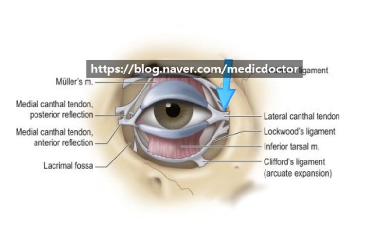
The lateral canthal ligament is also known as the lateral canthal tendon, and its location is indicated by the arrow in the image above.
If this area is injured, the lateral canthal ligament can become detached, leading to exposure of the conjunctiva.
Furthermore, the eye may not close completely, and the patient might appear to have a perpetually sad expression.
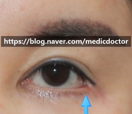
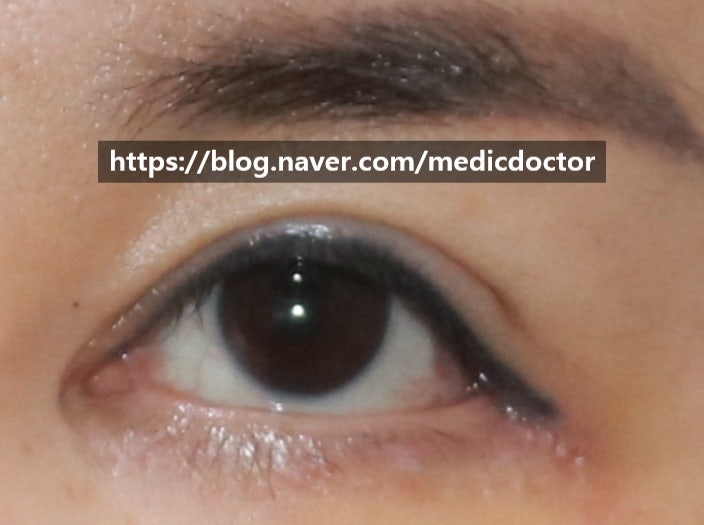
These are images from before surgery and 1 week after surgery, following suture removal.
The video below provides a clearer view:
The surgical principles are as follows:
1. Thorough removal of existing scar tissue.
2. Locating the detached lateral canthal ligament and restoring it to its original position.
3. Re-suturing to minimize visible scarring.
Is it just my impression that the eyes don't look smaller, but rather larger? ^^
The video conveys the severity of the issue more effectively than the photos.
Rather than undergoing indiscriminate lateral canthoplasty, it is better to opt for a moderate level of surgery that minimizes scarring.
I recommend a moderate lateral canthoplasty rather than undergoing revision surgery after an initial procedure.
Frequently Asked Questions
What are the side effects of excessive lateral canthoplasty?
Yes, damage to the lateral canthal ligament is a common side effect. If the ligament is damaged, the outer corner of the eye can become 'ㄷ'-shaped, and the conjunctiva may be exposed. There is also a risk that the eye may not close completely or may appear sad (a 'weeping' look).
What is the principle behind lateral canthoplasty revision surgery?
The principle is to remove existing scars and restore the ligament. First, the scars from the previous surgery are cleanly removed, and then the detached lateral canthal ligament is located and returned to its original, normal position. Afterward, it is meticulously re-sutured to minimize visible scarring.
When are the stitches removed after lateral canthoplasty reconstruction surgery?
The stitches are usually removed one week after the surgery. The text also compares the progress before surgery and one week after stitch removal, and from this point, the recovery process gradually becomes more natural.
Will my eyes become smaller after lateral canthoplasty revision?
No, your eyes do not necessarily become smaller. By restoring the damaged ligament to its normal position and correcting the structure, you may even feel that your eyes have become larger. It is important to perform the surgery at an appropriate level rather than excessive canthoplasty.