2026-05-29
Multiple Eyelid Folds and Failed Revision Surgery Case Study
A case study of a patient with multiple eyelid folds and severe scarring after three failed early revisions, successfully corrected through adhesion release.
Today, I would like to share a case study of a patient who visited for a follow-up one year after surgery.
Multiple eyelid folds, triple eyelids, and failed revision surgery case study
This is how I would define this case.
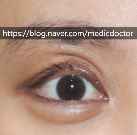
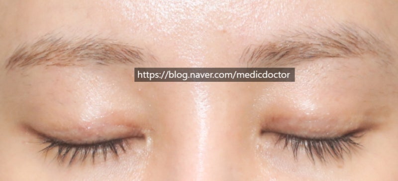
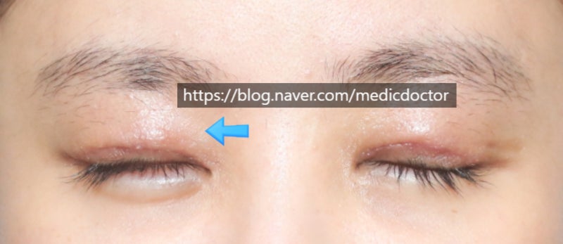
This is a photo of one of the patient’s eyes. The mucous membrane is severely exposed (eyelid eversion), and there are multiple folds and triple eyelids above the main eyelid crease.
This was not limited to just one side; it affected both eyes.
In this case, the patient had undergone surgery two months before visiting our clinic, and during that interval, they had already undergone three early correction attempts at the same hospital. They visited us seeking another early correction.
Actually, the ‘golden time’ for early correction is within two weeks post-surgery. While we typically do not perform early correction at the two-month mark, this patient was deeply distressed because their eyes had been operated on multiple times in such a short period.
However, I tend to actively recommend early correction in cases where daily social life is impossible, multiple folds occur, or there is a significant difference in eye size.
If it is merely a matter of slight asymmetry, it is better to wait six months.
Let’s look at the video first. This is the appearance before the surgery. It looks much more severe than in the photos.
During the surgery, the right side had a certain amount of fat and normal tissue remaining, but the patient’s left eye had almost no fat tissue and was in a serious state due to scar tissue.
This is the appearance immediately after surgery. Right after the procedure, the eyes do not close completely due to the effect of the anesthesia.
The core of the surgery is to release all existing adhesions, place the tissue back in its original position, and then ensure that re-adhesion does not occur.
It is crucial to prevent re-adhesion; if re-adhesion occurs, the surgery fails.
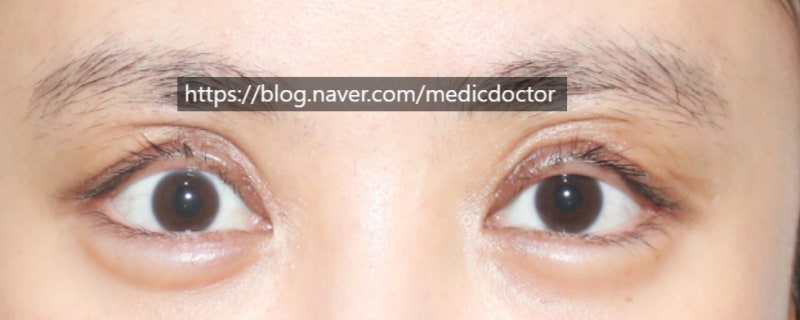
This is the appearance one week after surgery.
The right eye (left side of the photo) had some normal tissue left, so the shape looked good from the start. However, the left eye (right side of the photo) did not have much normal tissue, and because the tissue below the incision line was harder than the upper part, the upper area appeared relatively folded.
This is not a wrinkle caused by adhesion, but rather occurs because there is no volume in the area above the incision line. It resolves naturally once the swelling in the tissue below the incision line subsides.
You can trust the process and wait for this part to heal.
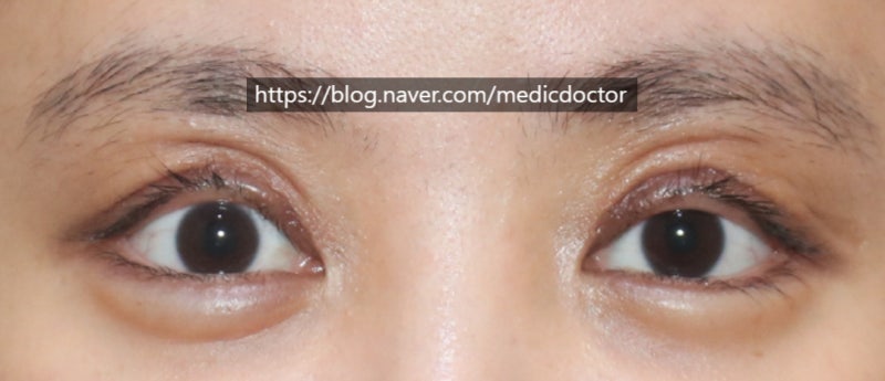
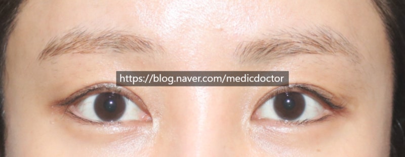
These are the photos from today’s visit.
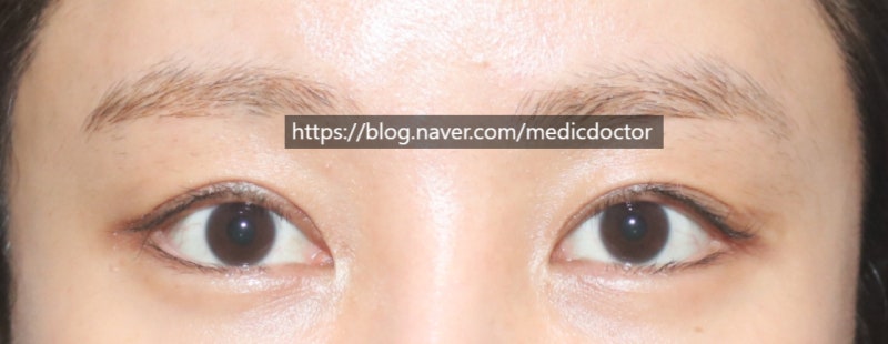
This is 10 months post-surgery. There has been a significant improvement. The only regret is that because too much skin was excised during the previous surgeries, further minute corrections may be difficult.
Scarring at the time of visit (the incision lines on both sides are asymmetrical). However, the distance from the incision line to the eyebrow is much closer on one side. Much more skin was cut away on the right eye (left side of the photo).
The photo taken today (10 months post-surgery) shows that the scars have also improved significantly. Some people mistakenly believe that a ‘step’ or ledge always forms after a ‘double-line release’ (lowering the crease), but that usually happens when the line is lowered significantly, when there is excess skin, or when existing scarring is severe. In this patient’s case, it did not occur because there was a lack of skin to begin with.
I am very relieved that the early correction went well and the overall surgical outcome is good.
I would also like to thank the patient for trusting me and waiting through the recovery process.
Frequently Asked Questions
When is the golden time for early correction of eye reoperation?
Yes, the golden time for early correction is usually within 2 weeks after surgery. However, if daily life is impossible due to discomfort, double eyelids have occurred, or the size of both eyes is severely different, early correction can be actively considered even after 2 months.
Should early correction be done even in cases of simple asymmetry?
No, if there is only simple double eyelid asymmetry, surgery is not performed immediately. It is safest and produces the best results to wait at least 6 months for the tissue to stabilize before proceeding with reoperation.
What is the core principle of double eyelid fold reoperation?
It is most important to release all existing incorrect adhesions. After releasing the adhesions, the key to successful surgery is to properly place the normal tissue in its original position and prevent readhesion. If readhesion occurs, the surgery will fail.
One week after surgery, the area above the incision line appears folded. Is this a side effect?
No, this may be a temporary phenomenon due to swelling and volume difference. It occurs when the tissue below the incision line is firmly swollen and the upper part lacks volume, and it will naturally resolve as the swelling below subsides, so you can wait with peace of mind.
Does a step (staircase phenomenon) always occur after a double-line revision surgery?
No, a step does not occur in all cases. It mainly occurs when the line is lowered significantly, when there is excess skin remaining, or when the existing scar is severe. This step phenomenon does not occur often in patients with insufficient skin.