2026-05-29
Understanding the Premaxillary Space: Essential Knowledge for Under-Eye Fat Repositioning
A guide to the anatomical structures of the premaxillary space, crucial for safe and effective under-eye fat repositioning surgery.
When performing under-eye fat repositioning surgery, a thorough understanding of the basic anatomical structures is essential to avoid surgical risks and prevent complications.
Please refer to the following details if needed:
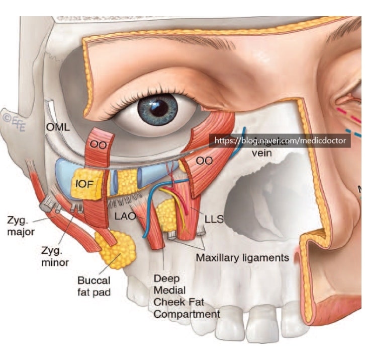
- Upper half roof: Orbital part of OOM
- Lower half roof: Mid-cheek SMAS
- Floor: LLS (levator labii superioris)
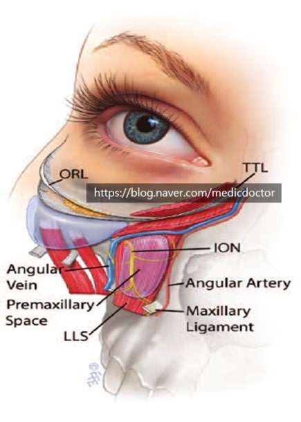
- Superior: TTL (tear trough ligament)
- Inferior: ML (maxillary ligament)
- Medial: Nasal side wall, LLSA (levator labii superioris alaeque nasi), Nasalis
- Lateral: Medial pupil line
This is a very well-known anatomical diagram.
During dissection, one must be careful of the angular vein and properly dissect the premaxillary space to lift the SOOF (Sub-Orbicularis Oculi Fat).