2026-05-29
Understanding the Premaxillary Space: Essential Knowledge for Under-Eye Fat Repositioning
A guide to the anatomical structures of the premaxillary space, crucial for safe and effective under-eye fat repositioning surgery.
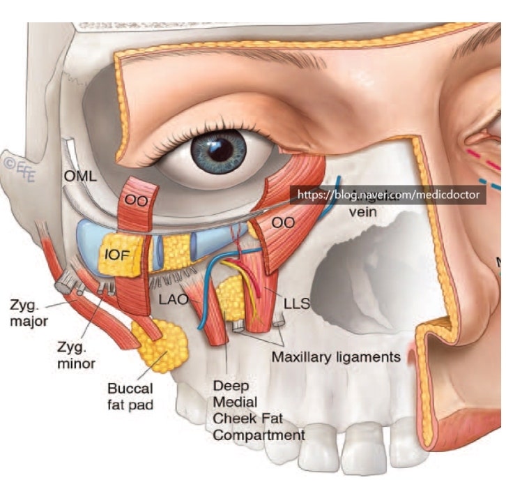
When performing under-eye fat repositioning surgery, a thorough understanding of the basic anatomical structures is essential to avoid surgical risks and prevent complications.
Please refer to the following details if needed:
- Upper half roof: Orbital part of OOM
- Lower half roof: Mid-cheek SMAS
- Floor: LLS (levator labii superioris)
- Superior: TTL (tear trough ligament)
- Inferior: ML (maxillary ligament)
- Medial: Nasal side wall, LLSA (levator labii superioris alaeque nasi), Nasalis
- Lateral: Medial pupil line
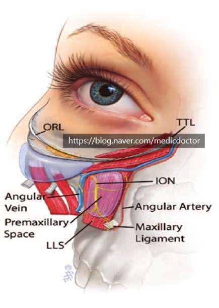
This is a very well-known anatomical diagram.
During dissection, one must be careful of the angular vein and properly dissect the premaxillary space to lift the SOOF (Sub-Orbicularis Oculi Fat).
Frequently Asked Questions
Why is it important to understand the anatomical structure during lower blepharoplasty?
It is important to avoid dangerous situations and prevent complications during surgery. Only with an accurate understanding of the complex anatomical structures around the eyes can a safe and successful lower blepharoplasty be performed.
Are there any blood vessels that require special attention during the dissection process?
Yes, the angular vein requires particular caution. Carefully avoiding damage to this blood vessel during tissue dissection for lower blepharoplasty is one of the critical points of the surgery.
How should the SOOF be handled during lower blepharoplasty?
The premaxillary space should be meticulously dissected and then lifted upwards. Properly dissecting this space and adequately elevating the SOOF is a key process in lower blepharoplasty.
What is the superior border of the premaxillary space?
The tear trough ligament (TTL) forms the superior border. According to the anatomical information in the text, the superior boundary is defined as the tear trough ligament, which serves as an important surgical landmark.