2026-05-29
Multiple Eyelid Folds, Mucosal Exposure, Eyelash Eversion, Ectropion, and Asymmetric Ptosis Correction (Converting Out-fold to In-out fold)
A comprehensive guide to correcting complex eyelid issues including multiple folds, mucosal exposure (eyelash eversion), and asymmetric ptosis through revision surgery.
Multiple eyelid folds, mucosal exposure surgery, eyelash eversion correction, ectropion, asymmetric ptosis, feat. (Resolving multiple creases and changing out-fold to in-fold)
Today’s topic is inspired by a patient who recently came in for a 4-month follow-up after surgery.
This patient had previously undergone an incisional ptosis correction at another clinic, which left severe scarring, significant mucosal exposure, and asymmetry.
The scarring is more visible under darker lighting conditions.
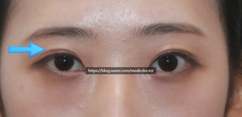
Looking at the patient’s left eye (right side of the photo) after their previous ptosis correction:
There was a depressed scar near the Mongolian fold caused by a forced attempt to create an out-fold, along with severe multiple creases and mucosal exposure.
The other eye appeared slightly sleepy. Under bright light, the eye-opening strength seemed significantly weak. While there is some underlying facial asymmetry, it can be further corrected to the greatest extent possible.
The goals of the surgery were as follows:
- Lower the fold line.
- Correct mucosal exposure and eyelash eversion as much as possible.
- Change the out-fold to an in-out fold style.
- Achieve maximum symmetry of the pupils.
- Resolve the multiple creases.
To lower the line, I set a new lower fold and released the existing adhesions. Regarding the multiple creases, I personally prefer to avoid fat grafting whenever possible, so this procedure was performed without it.
While mucosal exposure is typically corrected during the process, it is not the primary focus. If the fixation is done weakly and at a lower position, most mucosal exposure is corrected naturally. However, because aggressive correction can lead to functional side effects, I do not make mucosal and eyelash eversion correction the main objective.
I used the ‘dual-line’ technique (lowering the fold by creating a new line) to change the out-fold into an in-out fold.
Upon opening the patient’s right eye (left side of the photo) in the operating room, I discovered the levator muscle was detached and damaged, so I carefully performed the ptosis correction again. The patient mentioned that their right eye had been naturally smaller since birth.
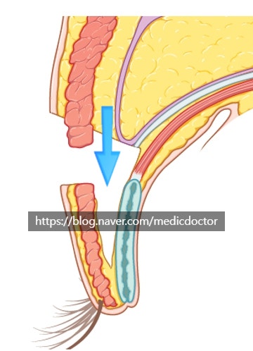
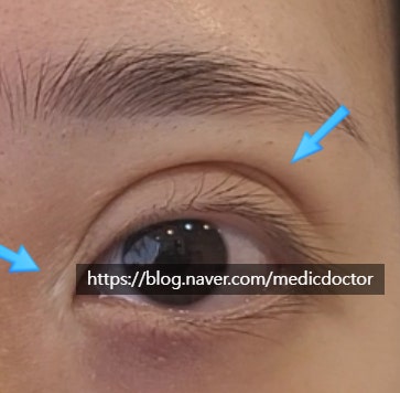
The diagram above is excerpted from Gunja Publishing (Aesthetic Plastic Surgery, Vol. 2).
The tissue under the eyelid is carefully dissected and fixed lower. However, excessive dissection can damage the orbicularis oculi muscle (the muscle that closes the eye), which may worsen lagophthalmos (inability to close the eye fully).
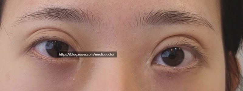
Appearance 4 months after surgery.
Due to skeletal facial asymmetry, the patient’s right side still appears slightly smaller. Because more ptosis correction was performed on that side, the eye is closer to the eyebrow, but this is the method that makes them look most similar. Most people would not notice the difference.
The surgery was successful thanks to the patient’s trust.
I also worked to release adhesions around the epicanthoplasty scars to minimize the depressed appearance.
During consultations, I tell patients that I will do my absolute best, but I never guarantee a specific result. While I obviously perform the surgery with the utmost care, some patients ask:
‘Is this definitely going to be corrected? I will only do the surgery if you give me a firm answer.’
I do not give such definitive promises. There are many variables during surgery, and in ‘extreme’ revision cases, it is difficult to make the eye look as if it never had surgery. Furthermore, individual satisfaction levels vary. However, I believe that when I recommend a procedure, it generally leads to a good outcome.
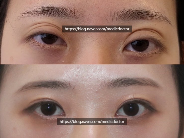
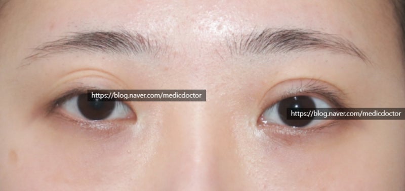
Before and after comparison.
Pre-operative video
Frequently Asked Questions
How are mucosal show and eyelash eversion corrected?
Yes, they are naturally corrected by adjusting the fixation to be low and weak. Attempting to correct mucosal show too aggressively can lead to functional side effects, so rather than making it the main focus of the surgery, we recommend improving it by releasing adhesions and applying appropriate fixation.
Is fat grafting always necessary to eliminate multiple eyelid folds?
No, it is not always necessary. As shown in the examples in the main text, multiple eyelid folds can be effectively resolved simply by meticulously releasing existing adhesions, without the need for fat grafting. Depending on the patient's condition, some prefer to proceed without fat grafting.
Can an out-fold double eyelid be changed to an in-out fold?
Yes, it is possible. By applying the double-line revision technique to lower the existing double eyelid line, an artificially created out-fold double eyelid can be changed to a natural in-out fold. During this process, depressed scars near the epicanthal fold are also improved.
Can asymmetrical ptosis that occurred after ptosis correction also be reoperated?
Yes, symmetry can be achieved through revision surgery. In cases where the eye-opening muscle is separated or damaged from a previous surgery, causing sleepy eyes, the muscle condition is checked in the operating room, and ptosis correction is meticulously performed again to maximize the symmetry of both pupils.
Is the result perfectly guaranteed for extreme eye revision surgery?
No, it is difficult to 100% confirm or guarantee the surgical results. Extreme eye revision surgery involves various variables, and it is challenging to achieve a perfect result like an unoperated eye. However, most patients can achieve good results if the surgery is performed with the utmost effort.