2026-05-29
Structure of Orbital Fat (Double Eyelid Fat Structure) Lower Eyelid Anatomy - Part 1
An in-depth look at the anatomical structure of the lower eyelid, focusing on the orbital septum, capsulopalpebral fascia, and arcuate expansion.
Structure of Orbital Fat (Double Eyelid Fat Structure; Orbital fat compartment and preaponeurotic fat)
1. Arcus marginalis origin (thickened by 1–3 mm at the periosteum, periorbita, and rim where fusion occurs); not a single layer but consists of several thin membranes.
2. Joins with the CPF approximately 5–6 mm below the lower margin of the inferior tarsal plate.
3. Upper reinforced portion by the CPF, lower unreinforced position not supported by the CPF.
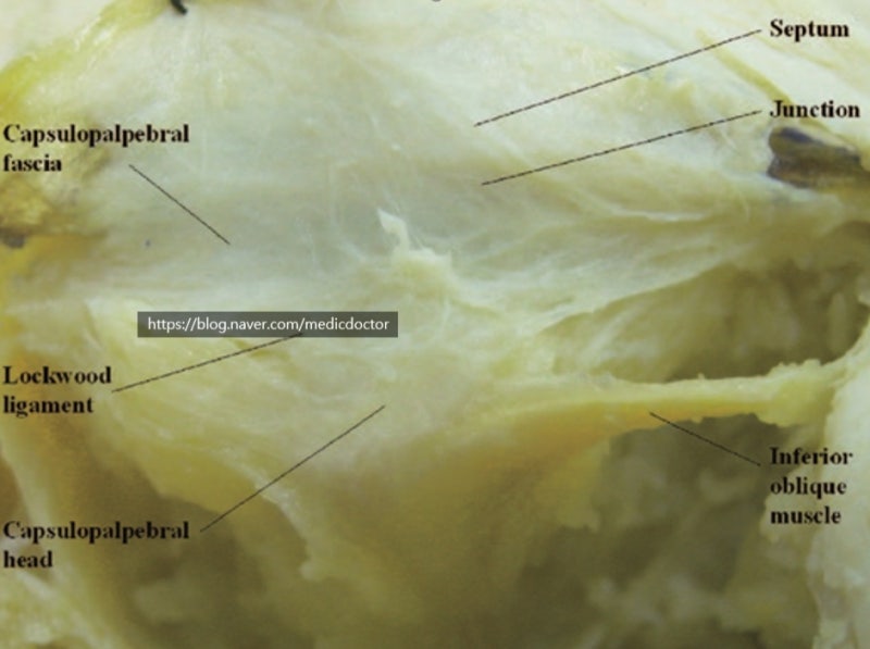
Capsulopalpebral fascia (CPF)
A well-defined connective tissue layer that mechanically links the lower eyelid with the downward retractor apparatus of the globe.
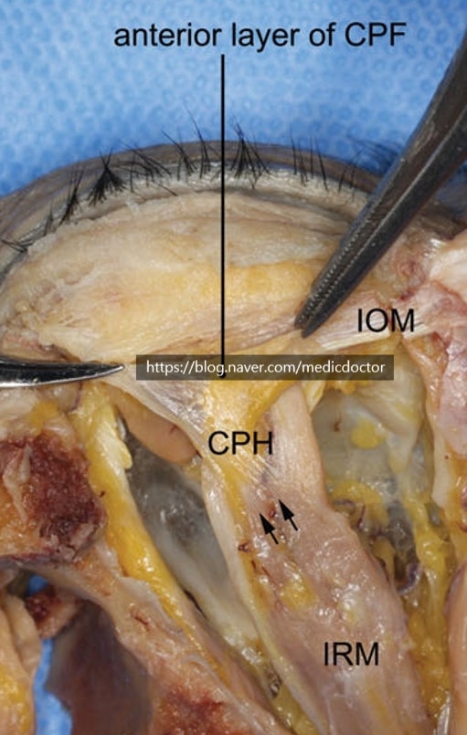
It is composed of two distinct layers that pass forward and upward from Lockwood’s ligament as dense fibrous sheets. The anterior layer (superficial layer) is coarse and inserts onto the orbital septum and deep fascia of the OOM. The dense posterior layer inserts onto the inferior border of the tarsal plate.
1) CPH (Capsulopalpebral head): Originates from the inferior rectus muscle fascia, wraps around the inferior oblique muscle, and reaches Lockwood’s ligament. Approximately 7 mm in thickness.
2) CPF: Courses from Lockwood’s ligament to the lower margin of the tarsus and subcutaneous tissue.
3) White glistening structure with two layers: an anterior thin layer and a posterior thick layer.
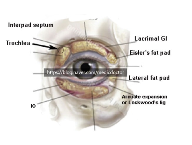
Arcuate expansion:
1) A fibrous band expanding from the infra-orbital rim to the medial canthal tendon. It is fan-shaped and tapered before attaching to the medial canthal tendon, with a width of 2–3 mm.
2) Located deep to the orbital septum and superficial to the inferior oblique muscle.
3) Overlaps the middle part of the inferior oblique muscle as it crosses and connects to the CPF.
4) The main Lockwood’s ligament and the arcuate expansion originate medially from the posterior lacrimal crest, dividing the central and lateral fat pads.
Frequently Asked Questions
Is the orbital septum a single membrane?
No, it is not a single membrane but is composed of multiple thin layers. It has a complex anatomical structure originating from the periosteum and orbital periosteum, connecting with the capsulopalpebral fascia (CPF) 5-6mm below the inferior border of the lower eyelid tarsus.
What is the role of the capsulopalpebral fascia (CPF)?
It is a distinct connective tissue layer that mechanically connects the lower eyelid and the downward traction system of the eyeball. It originates from Lockwood's ligament and runs anteriorly and superiorly, consisting of two distinct layers: a coarse anterior layer and a dense posterior layer.
What is the exact location of the arcuate expansion?
It is located deep within the orbital septum, situated on the surface of the inferior oblique muscle. It is a fibrous band extending from the inferior orbital rim to the medial canthal tendon, approximately 2-3mm wide, covering the middle portion of the inferior oblique muscle.
What are the anatomical criteria that divide the central and lateral fat pads of the eye?
The main Lockwood's ligament and the arcuate expansion are the criteria. These two structures originate medially from the posterior lacrimal crest and serve to clearly anatomically divide the central and lateral fat pads of the eye.