2026-05-29
눈의 지방 의 구조 (쌍꺼풀 지방의 구조) 하안검 해부학 -1.
눈의 지방의 구조 (쌍꺼풀 지방의 구조 ; Orbital fat compartment and preaponeurotic fat)<Lower eyelid>Orbital septum1. Arcus marginalis origin (periosteum, periorbita와 rim에서 1~3mm thickening되어 Fusion), not a si
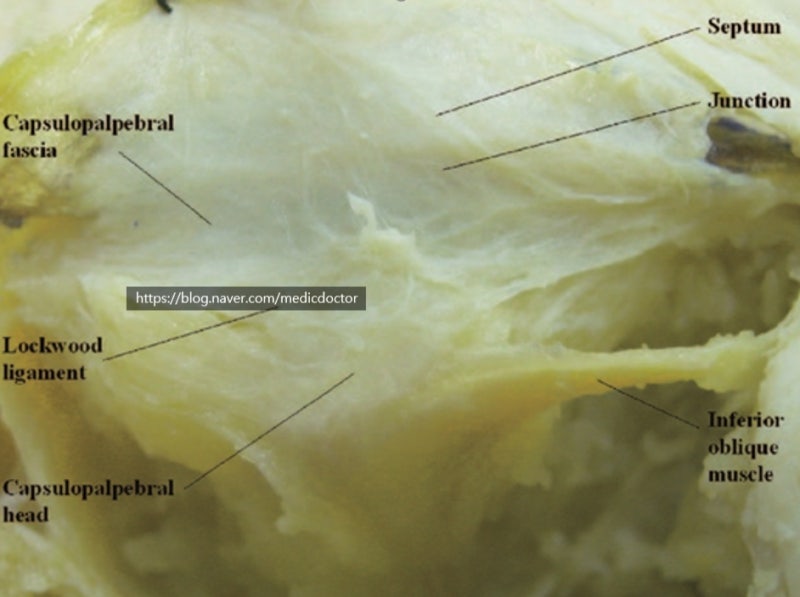
눈의 지방의 구조 (쌍꺼풀 지방의 구조 ; Orbital fat compartment and preaponeurotic fat)
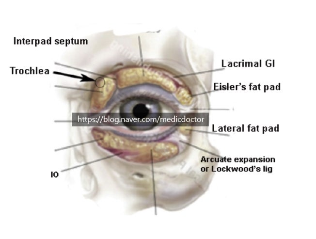
Orbital septum
1. Arcus marginalis origin (periosteum, periorbita와 rim에서 1~3mm thickening되어 Fusion), not a single layer but consist of several thin membrane
2. Inferior tarsal plate lower margin 아래 5~6 mm 아래에서 CPF와 join
3. Upper reinforced protion by CPF, lower unreinforced position not supported by CPF
Capsulopalpebral fascia
A well defined connective tissue layer that mechanically links the lower eyelid with the downward retractor apparatus of the globe.
It is composed of two distinct layers that pass forward and upward from the Lockwood’s ligament as dense fibrous sheets. The anterior layer or superficial layer is coarse and inserts onto the orbital septum and deep fascia of OOM. The dense posterior layer insderts onto the inferior border of the tarsal plate.
1) CPH(Capsulopalpebral head): originate from the inferior rectus muscle fascia, wrap around the inferior oblique muscle and reaches Lockwood ligament. 7 mm thickness
2) CPF: coursing from Lockwood ligament to lower margin of tarsus and subcutaneous tissue
3) White glistening structure with two layers : anterior thin layer and posterior thick layer
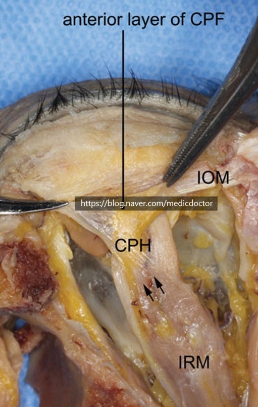
Arcuate expansion :
1) Fibrous band expanding from the infra-orbital rim to the medial canthal tendon
Fanshaped and tapered before attaching to the medial canthal tendon. 2~3 mm width
2) Located deep to the orbital septum and superficial to the inferior oblique muscle.
3) Overlapped the middle part of the inferior oblique muscle as it crossed and connected to the CPF.
4) Main Lockwood ligament와 arcuate expansion은 내측으로 posterior lacrimal crest에서 origin하여 central & lateral fat fad를 나눈다.
자주 묻는 질문
안와격막(Orbital septum)은 단일 막으로 이루어져 있나요?
아니요, 단일 막이 아닌 여러 겹의 얇은 막으로 구성되어 있습니다. 골막과 안와골막에서 기시하여 하안검판 하연 5~6mm 아래에서 안검판근막(CPF)과 연결되는 복합적인 해부학적 구조를 가집니다.
안검판근막(CPF)은 어떤 역할을 하는 조직인가요?
하안검과 안구의 하향 견인 장치를 기계적으로 연결해 주는 명확한 결합 조직 층입니다. 록우드 인대에서 시작해 앞쪽과 위쪽으로 주행하며, 거친 전방층과 촘촘한 후방층의 두 가지 뚜렷한 층으로 이루어져 있습니다.
궁상확장(Arcuate expansion)의 정확한 위치는 어디인가요?
안와격막의 깊은 곳에 위치하며, 하사근의 표면에 자리 잡고 있습니다. 하안와연에서 내안각건으로 확장되는 섬유 띠 형태로, 폭은 약 2~3mm 정도이며 하사근의 중간 부분을 덮고 있습니다.
눈의 중앙 지방과 외측 지방 패드를 나누는 해부학적 기준은 무엇인가요?
메인 록우드 인대와 궁상확장이 그 기준이 됩니다. 이 두 구조물은 내측으로 후방 눈물능선에서 기시하여 눈의 중앙 및 외측 지방 패드를 해부학적으로 명확하게 나누어 주는 역할을 합니다.
본 글은 강남서연성형외과의 원문(© 저작자)을 라이선스 계약 하에 재구성·편집한 콘텐츠입니다.
댓글 0